Zedu Weekly Wrap – 8 September 2023
We had a great coaching corner last night…
with a big turnout and some good discussions on early pregnancy loss. We’ll have the recording up on the website soon – but if you can’t wait for some excellent free ultrasound education check out the back catalogue – there’s something for everyone.
Things that sparked our interest this week include:
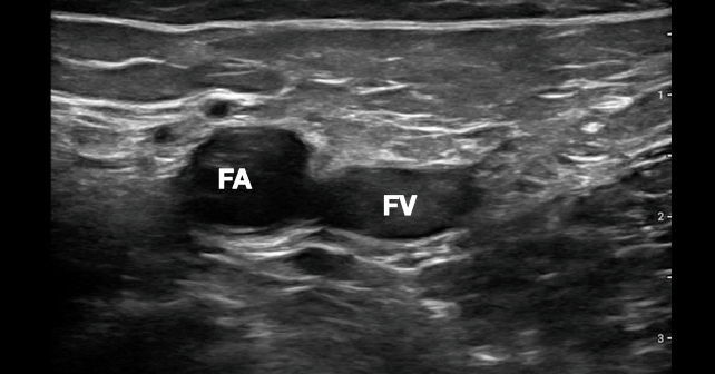
- a cool looking DIY Doppler ultrasound traning phantom that combines Lego, agarose gel and glycerin (5 September)
- an interesting piece by Nav Sidhu (@DrNavSidhu) on using gastric ultrasound to eliminate guesswork in periooperative airway management – page 115 of the Blue Book (8 September).
And don’t forget – if you have any news or ultrasound related research you’d like to highlight get in touch we’d love to hear from you.
Until this time next week…
Suean and Mike
Make 2024 your year of ultrasound and check out our range of scheduled and one-on-one options that will get you scanning like a pro.
Get the most out of your teaching moments – join our TRain the UltraSound Trainer (TRUST) course This isn’t a course on how to perform an ultrasound examination or use the technology – it’s designed to give you the teaching tools you’ll need to teach ultrasound on the run. In the words of a previous participant – it’s meta!
8 September
Check out the latest video from our Ultrasound Grand Rounds about Ultrasound and AI with @RubbleEM from @Yale_EUS https://t.co/8vrDbpe4wj#MetroEUS #Ultrasound #POCUS #ArtificialIntelligence pic.twitter.com/MougJWxMg1
— MetroHealth Emergency Ultrasound (@MH_EMultrasound) September 7, 2023
Throwback Thursday Case. 40s♀️hx HIV (last CD4 248)-1 month dyspnea. SC window first. VSS. Pox 93% RA #POCUS #FOAMed #FOAMcc #MedTwitter #MedEd @NephroP @ArgaizR @mount_md @msiuba @iceman_ex @TomJelic @cianmcdermott @curromir @IM_Crit_ @jminardi21 @jgutt @KalagaraHari @EM_RESUS pic.twitter.com/uWGlLL5red
— Robert Jones, DO (@RJonesSonoEM) September 7, 2023
Our narrative review on gastric ultrasound has just been published in @ANZCA Australasian Anaesthesia 2023.
Great to coincide with #ESRAWorld2023 pic.twitter.com/EKNVVSwZMs
— Dr Nav Sidhu (@DrNavSidhu) September 7, 2023
Click on the article title in list of contentshttps://t.co/67p227R5ET
— Dr Nav Sidhu (@DrNavSidhu) September 7, 2023
@ACEPNow
How To Safely Incorporate Ultrasound Into Cardiac Arrest Resuscitation
7 September
Next in my series on #POCUS in shock#FOAMed #FOAMus #echofirst @YouTube
Check it outhttps://t.co/XF29HBlZpV— Joseph Minardi (@jminardi21) September 6, 2023
6 September
A prenatal ultrasound report that notes a ‘short femur.’ What next? Our quick review of the recommended follow-up https://t.co/B7dorTZSOg #obstetrics #ObGynTwitter #foamOB #POCUS
— The ObG Project (@TheObGProject) September 5, 2023
The Game-Changing Use of Ultrasound on the Sideline: Revolutionizing Sports Medicine
5 September
What does #Lego, agarose gel & glycerin have in common?
Combined they create a Doppler #ultrasoundtraining phantom
Find out how:
Development and Evaluation of Doppler Ultrasound Training Phantom for Human Vessel Simulationhttps://t.co/2iNiIYyEUJ #POCUS #everythingisawesome pic.twitter.com/dVd0UgoQOS— Zedu Ultrasound Training (@zedunow) September 5, 2023
1/🧵Perioperative #POCUS or #EchoFirsst is valuable, but current methods are time-consuming.#AutoMAPSE on single-beat 3D #TEE assessed LV function with ⬆️feasibility & accuracy#Cardiotwitter #FOAMed #FOAMcc #MedTwitter
Paper📜:https://t.co/5LjPxfJZiM pic.twitter.com/lViCa4N1RT
— Anders Austlid Tasken (@AndersTasken) September 5, 2023
@AnatomyOrg

Radiology Case Reports
Imaging findings of fibroid torsion in pregnancy: A case report

4 September
Radiology Case Reports

3 September
@escardio

@escardio

2 September
@Applsci
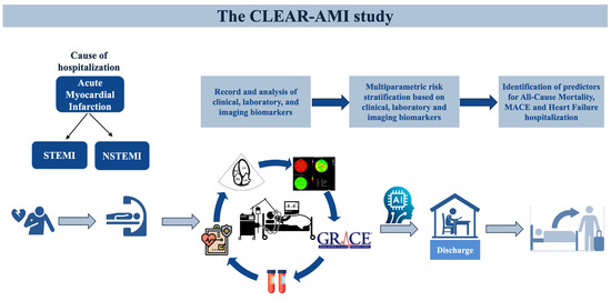
@Applsci
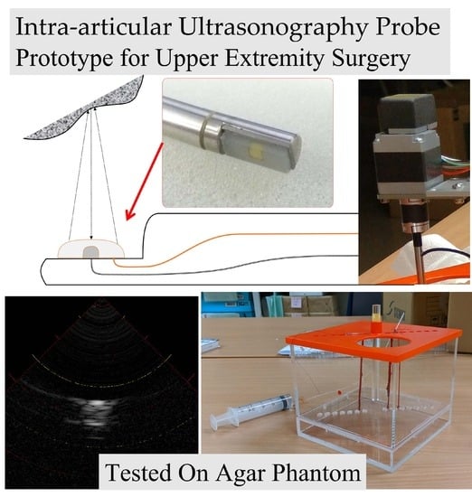
Academic Radiology @AURtweet

@ucsdsono
Case 32: Perforated Gallbladder

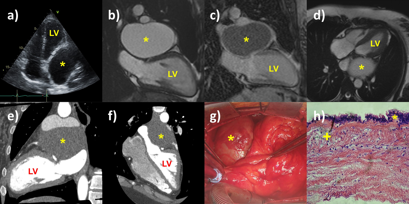
Echocardiography
“Bronchogenic cyst impersonating a pericardial lesion”: A case report
You may also like
Zedu Weekly Wrap – 9 October 2026
Zedu Weekly Wrap – 2 October 2026