Zedu Weekly Wrap – 18 February 2022
This week started with Valentine’s Day…
and highly appropriately we’ve been working through the intricacies of focused echo with a dedicated group of emergency physicians. A kind of week where “Roses are red, Doppler is too, but away is the only way signals turns blue”. Sorry for that, but we found that was the best way to steer away from the artery/vein red/blue relationship!
This week’s highlight is from Michael Prats and the Ultrasound GEL team talking a subject close to our hearts – Ectopic Pregnancy – Does POCUS Expedite Operative Management? Suean owes her life to POCUS and can attest to this from experience – check it out for yourself.
See you next week.
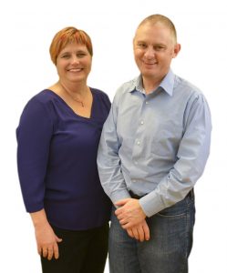
Mike and Suean
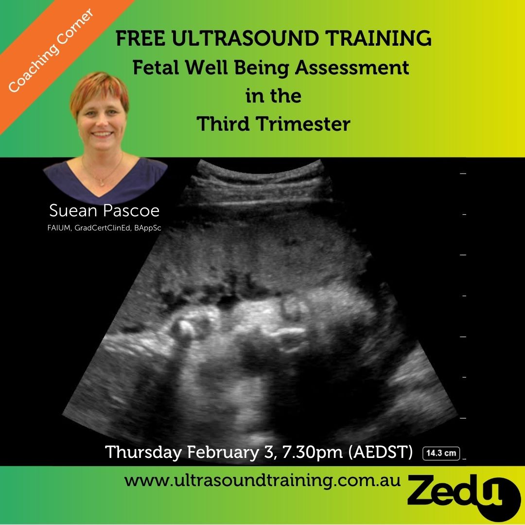
P.S. – The first Coaching Corner for 2022 – Suean’s discussion on fetal well being assessment in the third trimester – is now uploaded and available for free. Check out this fantastic resources and the entire the back catalogue here.
 18 February
— Stax_Baseball_Cards (@trigeminy_henry) February 17, 2022
Echocardiography Journal
Multimodality imaging of Abernethy malformation

@ACEPNow
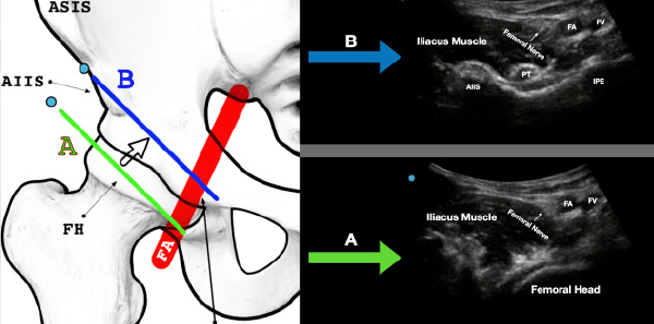
Benefits of Using the Pericapsular Nerve Group (PENG) Block
17 February
Detecting spontaneous retroperitoneal hemorrhage using a modified RUSH… https://t.co/m8rgU0Aj9w
— Zouheir Bitar (@zbitarsonoicu) February 16, 2022
Check out this week's #ultrasound discussion by @mtabbut about the Pediatric FAST exam in blunt abdominal trauma. #MetroEUS #POCUS #FOAMed #FOAMus #MedTwitter #EmergencyMedicine #Traumahttps://t.co/gH1ce317n3 pic.twitter.com/xbxBzanQ4b
— MetroHealth Emergency Ultrasound (@MH_EMultrasound) February 16, 2022
The #POCUS Consult: How Point of Care Ultrasound Helps Guide Medical Decision Making #FOAMED #FOAMUS @Wilkinsonjonny @zedunow @WINFOCUS @NephroP @POCUS_Society
👉 https://t.co/JU91hha9kk pic.twitter.com/ZIVthXon4D— Giuseppe Sfuncia (@Revivan75) February 16, 2022
and finally both images COMBINED (3/3)!
🗒️This is not a comprehensive list of everything but rather a mobile-friendly resource that can help at bedside#FOAMed #FOAMus@PittCardiology @SAEMAEUS @aroradrn @CanadianKayMD @nephrothaniel @AmrFBakarat @MasriAhmadMD @PACCMUS @PACCM pic.twitter.com/6xmmKYUUc4
— Mark Ramzy, DO, EMT-P (@MRamzyDO) February 16, 2022
I dagens avsnitt pratar vi POCUS med en av de stora i Sverige @chrismuhr.
Avnittet är i samarbete med GM Medical och @ButterflyNetInc
Finns där poddar finns.#pocus #ButterflyiQ #Foamed https://t.co/bay0KN0nTN— Arin Malkomian (@akuarma) February 17, 2022
Critical care #POCUS guided fluid resuscitation in septic shock: necessary? Which data?
💧superior vena cava diameter? inferior vena cava?
💧CVP?
Discussion ongoing…
🧫https://t.co/gw0arJQubtAuthors' reply to
🧫https://t.co/4sHJPJLANP🧫Original study https://t.co/YCQ397SK4I pic.twitter.com/lrzIdMTd1J
— Intensive Care Medicine (@yourICM) February 16, 2022
@ACEPNow @SonoStache
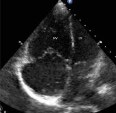
Preintubation Point-of-Care Echocardiography for Hemodynamic Optimization
16 February
@CasefromASE
Left Ventricular Metastases from Penile Squamous Cell Carcinoma

@PedRadJnl

15 February
US GEL PODCAST #120!
Ectopic Pregnancy – Expediting Operative Management? #POCUS #FOAMus #USGELhttps://t.co/E8EU36nFZr pic.twitter.com/JXZemnsIq0
— Michael I. Prats (@PratsEM) February 14, 2022
Appendix Gallery https://t.co/nxFF9zuH4B
— Stax_Baseball_Cards (@trigeminy_henry) February 14, 2022
Does fetal well being assessment in the 3rd trimester leave you baffled? Baffle no more – my #coachingcorner this month will give you confidence with a step-by-step approach
All #free #ultrasoundtraining for you – no gimmicks, no strings#POCUS #obstetrics #babies @zedunow https://t.co/gCN9JYYhxh— Suean Pascoe (@i_c_sound) February 15, 2022
Ultraschall Med
Visualized Urethral Mobility Profile During Urine Leakage in Supine and Standing Positions

@ASE360
HYPERSENSITIVITY CROSS-REACTIVITY FOR ULTRASOUND ENHANCING AGENTS AND COVID-19 VACCINES

@WINFOCUS
Pericardiocentesis: ultrasound guidance is essential

14 February
Before ordering morphine for an ankle reduction, consider a popliteal sciatic nerve block! Check out this article by Dr. Nazey Gulec & @MayaLinMD @NYPBrooklyn to learn more! #POCUS #FOAMed #FOAMus @NagdevArun @MGottliebMD @PennyLema @sonositu @javimedsimushttps://t.co/wYIk0h6uSw
— ACEP Ultrasound (@ACEP_EUS) February 13, 2022
13 February
This week's @VaveHealth CMO vid is just over seven years old & remains one of my teaching #meded favs. Anyone want to list the kind of effusion(s) seen here, the structure that helps us determine this, and why that structure looks like it is missing a lateral wall? #POCUS pic.twitter.com/ssy0RhcRJZ
— Renee Dversdal (@DRsonosRD) February 13, 2022
#POCUS Quizz:
What is that long structure running in the middle of the image?
'Right mid-axillary line with low frequency transducer'
1. Pulmonary vein
2. Pulmonary ligament
3. LVAD driveline
4. Artefact
5. I don't know #CardioTwitter #echofirst pic.twitter.com/fRKRKv9CYL— Hatem Soliman Aboumarie, FRCP, FEACVI, FASE, FHEA (@EchoSoliman) February 12, 2022
@CasefromASE

@AJOG_thegray

12 February
Thought provoking article on the ethical considerations and obligations for primary care to adopt a clinical skill #POCUS that has both an evidential basis for benefit and is practically attainable https://t.co/OTB18SrxyL @rnzcuc @opotikigp @ParmyDeol @krishkashap @drbarai pic.twitter.com/DmfYPEYrAN
— Kelvin Ward (@UCUltrasound) February 11, 2022
EM@3AM: Cellulitis #FOAMed @secohen11 @EMHighAK @long_brit https://t.co/ozmUdTdGrF
— emDOCs Team (@emdocsdotnet) February 12, 2022
Journal of Clinical Ultrasound

@CASEfromASE
A Primer on Three-Dimensional Transthoracic Echocardiographic Imaging of the Tricuspid Valve

@PLOSONE
@PLOSONE
@PLOSONE
@PLOSONE
@PLOSONE
The impact of skeletal muscle disuse on distinct echo intensity bands: A retrospective analysis
@PLOSONE
We have a new #CoSTR #ALS Systematic Review on Diagnostic Test Accuracy of Point-of-Care Ultrasound during CPR to indicate the etiology of cardiac arrest available for public comment !
⛓ Read CoSTR 👉 https://t.co/gKwWhqDhLF and leave your comment! pic.twitter.com/GgD43984j5— ILCOR (@Ilcor_org) February 10, 2022

You may also like
Zedu Weekly Wrap – 9 October 2026
Zedu Weekly Wrap – 2 October 2026