
Zedu Weekly Wrap – 23 December 2022
‘Tis two nights before Christmas…
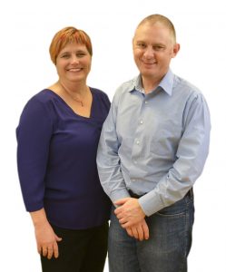
and if you’re anything like us there’s still a long list of things to do. When Saturday night arrives we’ll all collapse in a heap.
So will make this short. We’re closing down today, and returning on Monday 9 January. There’ll be no weekly wrap over that time, but we might sneak the odd look a the socials (internet connection allowing) when not at the beach.
Wishing everyone out there a safe and happy holiday season – may your probe stay clean, your gel warm and your battery charged. We hope you all get the opportunity to relax, recharge and catch up with family and friends. And to those working over this special time – a massive thank you.
Highlights for this week include new learning resources from Joseph Minardi (@jminardi21), a swag of articles from the Journal of Clinical Medicine (@JCM_MDPI) and the latest Ultrasound G.E.L. podcast from Michael Prats (@PratsEM).
That’s all for now – see you in 2023…
Happy holidays
Suean and Mike
P.S. – follow us on Mastodon – https://med-mastodon.com/@zedunow
P.P.S. – Catch up with all 30 episodes of the Coaching Corner – covering a wide gamut of ultrasound topics – and always free 🙂 The next instalment will be Thursday 2 February see you in 2023! (Let us know if you want something specific covered…)
PS – Join the POCUS Educators / Seed Support Network on Facebook – a safe space to share education tips, resources and issues. People from around the world are joining in so come on down.
Get the most out of your teaching moments – join our TRain the UltraSound Trainer (TRUST) course This isn’t a course on how to perform an ultrasound examination or use the technology – it’s designed to give you the teaching tools you’ll need to teach ultrasound on the run. In the words of a previous participant – it’s meta!
6 – 7 May 2023 | 7 – 8 October 2023
23 December
Urinothorax anyone? 🤔 Check out our newly published case report! #MedTwitter #pulmonology #POCUS https://t.co/LIkJ4jvLdG
— Luderve Rosier, MD (@LuderveRosier) December 22, 2022
New in JACEP Open from @EVMSedu: Hook effect in gestational trophoblastic disease: An emergency department case presentation. #FOAM, #Obstetrics, #EmergencyMedicine, #POCUS https://t.co/mCVo55WcPq @EmergencyDocs @ACEPNow @WileyHealth @wileyinresearch pic.twitter.com/qkzn9CgI8u
— JACEP Open (@JACEPOpen) December 22, 2022
Excited to add to evidence of use of #POCUS. A case report highlighting intra-arrest use of #POCUS assisting in decision making and leading to good outcome for the patient.
Plus more stuff on #DVT & #Pulmonaryembolism@tonyjoy81 @haythemjarad @awaischeema https://t.co/5bFJsEH1D5— Salman Naeem (@salmannaeem217) December 21, 2022
@JCM_MDPI
Contribution of Ultrasound in Current Practice for Managing Juvenile Idiopathic Arthritis

@JCM_MDPI
Fetal Tibial Artery Doppler in Late IUGR Fetuses: A Longitudinal Study

22 December
Want to get started recognizing left ventricular dysfunction? Cardiomyopathies? #echofirst #POCUS #FOAMed #FOAMus
Here's a good place to starthttps://t.co/X9jxkiLJRa— Joseph Minardi (@jminardi21) December 21, 2022
New Ultrasound of the Month Post – what do you see? Learn more with @jsobocin #FOAMed #FOAMus https://t.co/vaSOqshtDe @mingespg @martina_diazb @sonostolz pic.twitter.com/RonUk7XqNW
— UC EM Residency (@TamingtheSRU) December 21, 2022
@CASEfromASE ft @VLSorrellImages
Should Doppler Always Be Capitalized?

Radiology Case Reports

Radiology Case Reports
Male breast myofibroblastoma: Imaging features and ultrasound-guided core biopsy diagnosis

Radiology Case Reports

Radiology Case Reports
A rare case of left atrial myxofibromyosarcoma

Radiology Case Reports
Atrial flutter with slow ventricular response revealing a double discordance: A case report

Radiology Case Reports
Male breast: An unusual case of metastasis of squamous cell carcinoma of the skin

@JCM_MDPI
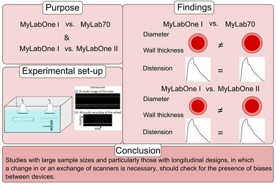
Detectable Bias between Vascular Ultrasound Echo-Tracking Systems: Relevance Depends on Application
21 December
Next #POCUS lecture out:
Echo for Fluid Responsiveness (FR)
Summary slides and key points ⤵️🧵@HarvardPulm @PCCSM_BIDMC @MGH_PCCM @MGHHeartHealth @MGHMedicine @mghmedres #MedTwitter #MedStudentTwitter #PCCMtwitter pic.twitter.com/LUjQVZeGmP— Jonah Rubin MD, FCCP (@JonahRubinMD) December 20, 2022
#Echocardiography is essential in diagnosing cardiogenic shock secondary to acute MR.
Read our @CASEfromASE article, Cardiogenic Shock Secondary to Acute Mitral Regurgitation With Nonischemic Etiology Successfully Stabilized by Transcatheter Intervention! https://t.co/Y6BcXJVmpC pic.twitter.com/5YW4SgwHuR
— American Society of Echocardiography (@ASE360) December 20, 2022
emDOCs Podcast – Episode 68: Tubo-Ovarian Abscess #FOAMed @EMHighAK @long_brit @MPrizzleER https://t.co/Uxt1VsHt7o
— emDOCs Team (@emdocsdotnet) December 20, 2022
@TheSDMS

@CJCJournals
A ring with endocarditis – an unfortunate marriage

@escardio
Systematic lung ultrasound in Omicron-type vs. wild-type COVID-19

@escardio

20 December
US GEL PODCAST #138!
Ultrasound vs Landmark Subclavians – Randomized + Controlled #FOAMus #FOAMcc #USGELhttps://t.co/iwaZ28hIlF pic.twitter.com/EaDdVBkbuL
— Michael I. Prats (@PratsEM) December 19, 2022
Next up in my series on heart and lung #POCUS
Left ventricular failureCheck it out #FOAMed #FOAMus #Echofirst @WVUMedSchool @WVUMedicine @WVUEmergencyMed @wvuemeducation @WVU_Ultrasound https://t.co/X9jxkiLJRa
— Joseph Minardi (@jminardi21) December 19, 2022
@PLOSOne
@JCM_MDPI
Bedside Ultrasound for Hemodynamic Monitoring in Cardiac Intensive Care Unit

Heart, Lung and Circulation

Annals of Emergency Medicine
Young Boy With Scrotal Swelling

Man with chest pain and dyspnea

Young man with nausea after a traffic collision


Man With Abdominal Pain and Malaise

Young Man With Abdominal Cramps and Bloody Stools


19 December
1/ Midline en Difficult Vein Access (DIVA)#pocus #nurse @coreultrasound @Sociedad_SEMI @SAEMAEUS @zedunow @AIUMultrasound @ACEP_EUS @SCUFellowships @UltrasoundJelly @WINFOCUS @EnfermeriaSEMES @ecoSEMES_ @EnfrmraSaturada @EcosemfyC @CGEnfermeria @soyenfermera @FenfUdelar pic.twitter.com/lWXbCOyJHG
— Yale Tung-Chen (@yaletung) December 18, 2022
@PLOSOne
18 December
@JCM_MDPI

@JCM_MDPI

17 December
@WFUMB

The Journal of Vascular Access @SAGE_Publishing

The Journal of Maternal-Fetal & Neonatal Medicine @tandfonline
Prenatal ultrasound evaluation of fetal cutaneous hemangioma and related complications
@ehj_ed
Artificial intelligence in cardiac imaging: where we are and what we want

You may also like
Zedu Weekly Wrap – 9 October 2026
Zedu Weekly Wrap – 2 October 2026