Zedu Weekly Wrap – 24 February 2023
Things are heating up here…
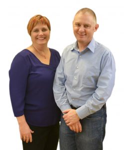
as Summer hits it’s peak, and scheduled courses run back to back with one-on-one and small group training in between. It’s great to see the place buzzing again after the covid hibernation. If you have training that was put on the backburner over the past two years get in touch – we’ll get you up and running in no time.
Plenty of free ultrasound and POCUS learnings to pick through this week. Highlights include nerve blocks for the ED from @MH_EMultrasound, an interesting piece on whole-body imaging using ultrasound tomography with 3D data acquisition (volography) from @AcadRadiol, and research on computer assisted analysis of lung ultrasound patterns that could assist both diagnosis and standardisation of illness severity scoring via @Crit_Care.
This and more – all worth checking out. And as always, if you have research, news, views or resources you’d like included in the weekly wrap please drop us a line – we are always up for an ultrasound chat.
See you next week.
Suean and Mike
P.S. Check out February’s Coaching Corner – the tips & tricks you can use to take the pain out of foreign bodies – available now and free on the website.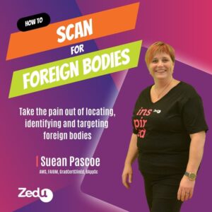
Get the most out of your teaching moments – join our TRain the UltraSound Trainer (TRUST) course This isn’t a course on how to perform an ultrasound examination or use the technology – it’s designed to give you the teaching tools you’ll need to teach ultrasound on the run. In the words of a previous participant – it’s meta!
6 – 7 May 2023 | 7 – 8 October 2023

24 February
Check out our Ultrasound Grand Rounds video by @KCaja_24 about Ultrasound Guided Nerve Blocks.https://t.co/nCPkGA8RaT#MetroEUS #POCUS #ultrasound #Trauma #NerveBlocks pic.twitter.com/CtttmmdjdN
— MetroHealth Emergency Ultrasound (@MH_EMultrasound) February 23, 2023
In this episode, @ABUltrasound & @miller_pocus chat with Exo’s VP of AI, @dornoosh, about #artificialintelligence in #healthcare. Dornoosh discusses bias in #AI, working to removing it, & more.
🎧Tune in: https://t.co/fVvIZXCVrt#Podcast #FOAMed #POCUS #Ultrasound #MedTwitter pic.twitter.com/GTX3XrhEmy
— Exo (@exoeffect) February 23, 2023
@EditorsVJEM

@amjmed

@JCM_MDPI
Sonographic Features of Onychopapilloma: A Single Center Retrospective Observational Study

@AcadRadiol
Whole-Body Imaging Using Low Frequency Transmission Ultrasound

23 February
Listening to "Is it safe to perform and teach lung ultrasound?"
You can hear in any podcast apphttps://t.co/tKCvVPuxJc@CmeNuvance @NuvancePOCUS @NephroP #LUS #Lungultrasound #POCUS #IMPOCUS
— Danburymedicalfiles (@Danburymedfiles) February 22, 2023
Sometimes the soft tissue surrounding fractures are so swollen that LM-based hematoma blocks don’t seem to work. Adding #POCUS to your procedure can help with precision placement of that anesthetic!
Check out the video for more info: https://t.co/HGwldBiWuo#FOAMed #MedEd pic.twitter.com/hUJn3mzpz4
— Jailyn/Ben (@coreultrasound) February 22, 2023
Flank pain? Creatinine elevation? Complicated UTI? Better #POCUS those kidneys
Learn how #FOAMed #FOAMus @wvuemeducation @WVUEmergencyMed @WVUMedicine @WVUMedSchool @WVU_Ultrasound @NephroP https://t.co/048SYxpnaF— Joseph Minardi (@jminardi21) February 22, 2023
22 February
Find that fracture in seconds (Or use it during reduction to ID adequate alignment) w/ #POCUS!
Check out the video for the whole technique. Also, how do YOU use ultrasound for bones? https://t.co/HGwldBiWuo#FOAMed #MedEd pic.twitter.com/cF7nD9aih3
— Jailyn/Ben (@coreultrasound) February 21, 2023
Musculoskeletal ultrasound tutorial – Anterior inferior tibiofibular ligament (AITFL) and syndesmosis.🎥https://t.co/ZY5dc8WUA1 pic.twitter.com/8pPx7B0Heo
— The Ultrasound Site – MSK ultrasound training (@mskultrasound) February 21, 2023
New Link with the latest update https://t.co/1Q9Um5EuJe
— Krunal H. Patel ⚕️ (@MDKrunalP) February 22, 2023
@Crit_Care

@JCM_MDPI

@J_Cardiol

@EMNews
The Speed of Sound – Symptom: Abdominal Pain

@MDPIOpenAccess
OCTA and Doppler Ultrasound in Primary Open-Angle Glaucoma and Normal-Tension Glaucoma

21 February
Check out Episode 6 of #BlockItLikeItsHot to hear @amit_pawa & @jeffgadsden talk about Blocks for Breast surgery!
In this snippet they talk about @Ropivacaine ‘s paper here https://t.co/6JkX2WujjA pic.twitter.com/9NdpAVAz2j
— Block It Like It’s Hot Podcast (@BlockIt_Hot_Pod) February 20, 2023
🚨🚨🚨🚨another edition of #fromthetrenches🚨🚨🚨🚨
Recent case of mine —> clinical details altered for anonymity
59 year old male presents to ED with SOB. Family states was at home at fell in bathroom. Hx of HTN/DM. Unsure of meds or compliance to therapy. Arrival BP 79/42 😱😱— nick pulmcrit (@pulmtoilet) February 21, 2023
@ThoraxBMJ
Rare cause of massive haemoptysis diagnosed by ultrasonography in the ICU

@JCM_MDPI

@AnnalsofEM

@AnnalsofEM

@AnnalsofEM
Sudden Ankle Pain in a Healthy Young Man

@AnnalsofEM

@AnnalsofEM
Woman with Acute Finger Pain and Swelling

@AnnalsofEM

20 February
@SciReports

19 February
@JCM_MDPI

18 February
Pediatric Cardiology

@WFUMB

@MDPIOpenAccess
The year 2021 in the European Heart Journal: Cardiovascular Imaging Part II

@ASE360

@CASEfromASE

@CUREUS

Neonatology
European Consensus Guidelines on the Management of Respiratory Distress Syndrome: 2022 Update
You may also like
Zedu Weekly Wrap – 9 October 2026
Zedu Weekly Wrap – 2 October 2026