Zedu Weekly Wrap – 25 August 2023
We’ve had a great week this week…
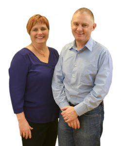
getting in depth on all things baby with our comprehensive obstetric program. Lots of new mums eager to see their bub, and docs eager to scan them. A match made in ultrasound training heaven! And a lot of families have sent us through pics of their newest family members, so we’ve started to catalogue them on our new zedupregnancyscans Insta account – be sure to check it out for a dose of cuteness 🙂
Not so cute (I guess it depends on who you are) but equally captivating is the latest in ultrasound goodies this week – check out:
- Metro Health Emergency Medicine (@MH_EMultrasound) and their video on MSK ultrasound in emergency medicine (24 August)
- a piece by the team at Saint Emlyns (stemlyns) part 1 of an intro to paediatric ultrasound in the ED (23 August).
Plus there’s a whole range of other #FOAMed resources to catch up on.
The weekend’s calling – better get to mowing the lawn!
See y’all next week.
Suean and Mike
Make 2024 your year of ultrasound and check out our range of scheduled and one-on-one options that will get you scanning like a pro.

Get the most out of your teaching moments – join our TRain the UltraSound Trainer (TRUST) course This isn’t a course on how to perform an ultrasound examination or use the technology – it’s designed to give you the teaching tools you’ll need to teach ultrasound on the run. In the words of a previous participant – it’s meta!
25 August
@ISUOG

24 August
This week highlights #ultrasound guided NBs in the ED (Brown et. al.)
Written for a USA audience the benefits of USGNB for pain mgmt are universal.
Are you using NBs in the ED? Have you faced challenges?Join the conversationhttps://t.co/pz9ksVdBjX#POCUS #ultrasoundtraining pic.twitter.com/DsU15gae8T
— Zedu Ultrasound Training (@zedunow) August 24, 2023
Check out our latest video from our Ultrasound Grand Rounds about MSK Ultrasound in Emergency Medicine with Sports Med Dr. Barnes and Dr. Raineyhttps://t.co/RoMXt6begw#metroEUS #POCUS #ultrasound pic.twitter.com/g8h6ttmCZa
— MetroHealth Emergency Ultrasound (@MH_EMultrasound) August 23, 2023
Getting a pocket #ultrasound might seem like a great idea, but Yash Chavda, DO, says #handheld #US presents problems, including privacy concerns & expensive upgrades. #POCUS #FOAMUS https://t.co/wuR8cRc53K pic.twitter.com/55VWrC0lGE
— Emergency Medicine News (@EMNews) August 23, 2023
Ultrasound can dramatically increase the success of diagnosis of a wrist effusion as well as assist in an efficient and accurate aspiration/injection of that joint. Check out the latest 5MS video to learn how!https://t.co/40tAwzFPd2#FOAMed #MedEd #FOAMus pic.twitter.com/G9ATYOa9AZ
— Jailyn/Ben (@coreultrasound) August 23, 2023
@escardio

@escardio

23 August
Part 1 of an introduction to paediatric ultrasound in the emergency department from Dr Pete Hulme @Dr_Pete_EmMed @stemlyns #FOAMedhttps://t.co/FZyL1pzXPF
— Saint Emlyn's (@stemlyns) August 23, 2023
@escardio
Severe aortic stenosis detection by deep learning applied to echocardiography

22 August
A new study about serial point-of-care ultrasound, which can stabilise patients faster!!! https://t.co/t1SbvjM808 #PoCUS #US #ultrasound #emergency #emergencymedicine #research
— Michael Dan Arvig (@Doktorarvig) August 21, 2023
@ASE360

@JCM_MDPI
21 August
Journal of Clinical Ultrasound
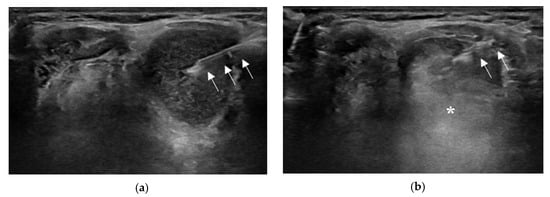
Ultrasound features and differential diagnosis for superficial nodular fasciitis

@WINFOCUS
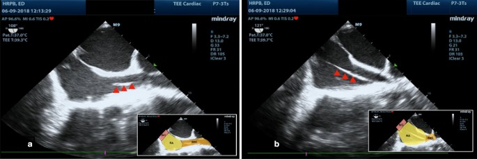
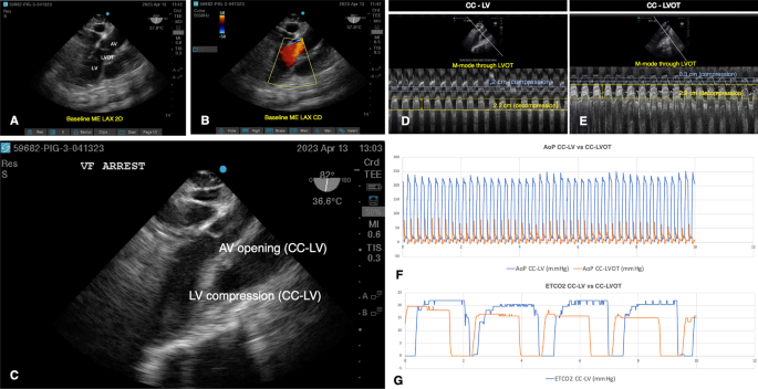
Transesophageal echocardiography (TEE)-guided transvenous pacing (TVP) in emergency department
@WINFOCUS
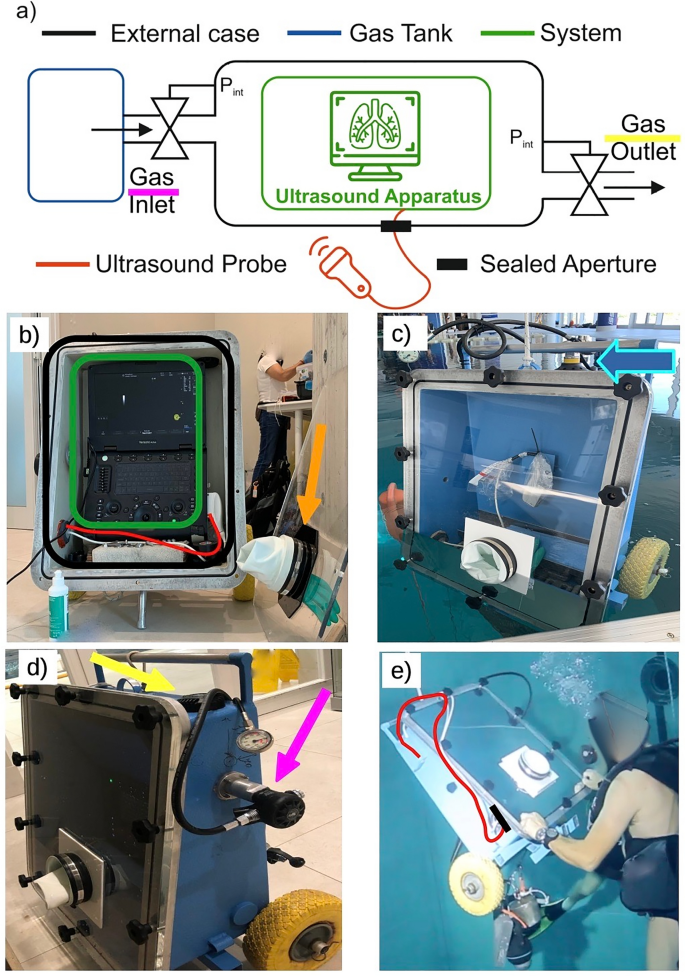
Feasibility of chest ultrasound up to 42 m underwater
20 August
19 August
Little study on how long it typically takes to perform a PoCUS examination (spoiler alert—not long!!). Congrats David, Xenia, Caroline, and Bowen! https://t.co/KtooBcLr6I pic.twitter.com/oex9AusKEv
— Meghan Herbst (@EUSmkh) August 18, 2023
@Crit_Care
@PLOSOne
You may also like
Zedu Weekly Wrap – 9 October 2026
Zedu Weekly Wrap – 2 October 2026